From the Journal of the American Orthodontic Society · March/April 2013
Dual Arch Orthopedics: Phase I Treatment in the Early Mixed Dentition
Journal of the American Orthodontic Society, March/April 2013, pp. 36–40
The debate over early orthodontics and/or orthopedic arch development in the growing child has long been controversial. There are numerous articles in the literature in favor of early treatment strategies beginning in the mixed dentition (1–15). It makes sense to correct problems early rather than let them linger and even worsen over time. I can think of no other area in medicine that allows problems to deteriorate over time without intervention. In order to provide treatment early, however, it is necessary to identify developing problems early, or at least to anticipate these problems before they occur so that corrections can be made in a timely and efficient manner. The American Association of Orthodontists (AAO) recommends children get an orthodontic screening by age 7. The timing of this recommendation, by no accident, coincides with exchange of the front teeth (early mixed dentition). Many an experienced clinician has heard the all too familiar question from a concerned parent whose child is at this stage, "will my child need braces?" In the interest of doing what is best for our patients, we should as dentists, be able to respond correctly, and provide the necessary treatment guidance at this stage, or be able to refer the child to someone who can treat appropriately. Watchful waiting does the child no good and may even cause more harm in the long run. The window of opportunity demands diligence on the part of the treating dentist, as many patients will not achieve proper facial balance, esthetics, or optimal health without early intervention.
Numerous factors are implicated in the literature as to the causes or etiologies of malocclusion ranging from habits, aberrant swallow patterns, obstructed nasopharyngeal airways (7, 16–20), rest oral posture (21), modern diets (22, 23), environmental causes (22) to natural birth (24). Growth is rapid in young children, so orthopedic corrections need to be made early to prevent undesirable patterns that are less correctable later. Orthopedic changes are limited in older children where facial growth is near completion. Experience in dealing with growing children has shown me that malocclusions develop early and worsen over time. By age twelve 89% of children have dental malocclusion (25). Thus, there is a critical need to diagnose and treat developing malocclusions in young children.
In this article I will introduce an early treatment system which I have developed and termed "Dual Arch Orthopedics". The concepts and treatment strategies are not new, and have been described previously (26, 27, 28). However, modifications and changes have been made so that I could apply these concepts to the dentition of the growing child. Dual arch orthopedics enhances growth and dental development so these can proceed without interference or alteration, as nature intended. The child can then grow up with a well balanced and attractive face, straight teeth, a beautiful smile, and healthy jaw joints.
Indications include:
- Narrow arch widths
- Anterior crowding and or rotations
- Ectopic eruptions of the lower incisors
- Early loss of a primary cuspid(s)
- Blocked out or rotated incisors noted on x-ray
- Resorption of adjacent tooth roots noted on x-ray
To demonstrate the dual arch orthopedic technique, I have selected a representative case, G.J., an 8 year 4 month Caucasian male in an early mixed dentition. Complete orthodontic records were taken on 11-18-08. Shortly after, a comprehensive orthodontic workup and diagnosis was completed which revealed a total space discrepancy of 3.5mm, and 5.5mm in the lower and upper arches respectively. There was an anterior cross bite involving the upper right lateral incisor, and a lower midline deviation to the left 1.0mm. The upper left lateral was rotated 90 degrees and was blocked out. Both lower lateral incisors were blocked from their normal eruption path due to an anterior arch constriction. Although the growth pattern was deemed normal, the facial profile revealed retrusion of both jaws. In addition, G.J. had thin lips and an obtuse nasio-labial angle. G.J. was referred to a pediatric allergist for evaluation and management of chronic allergies at the start of treatment. (Figs. 1a–c)
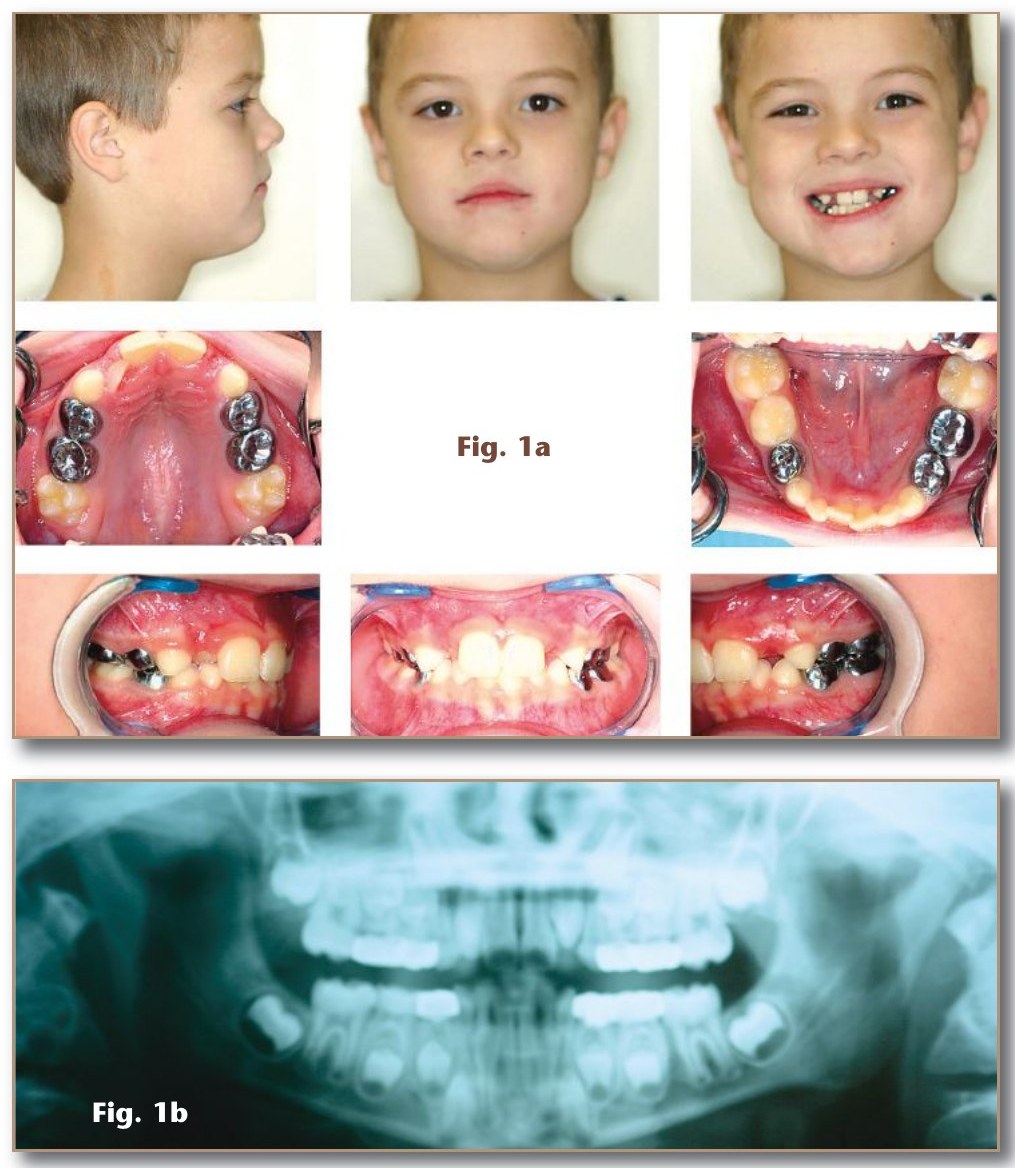
- Fig. 1a Pre-treatment facial photographs (profile, frontal, smiling), G.J., age 8y4m.
- Fig. 1b Pre-treatment panoramic radiograph.
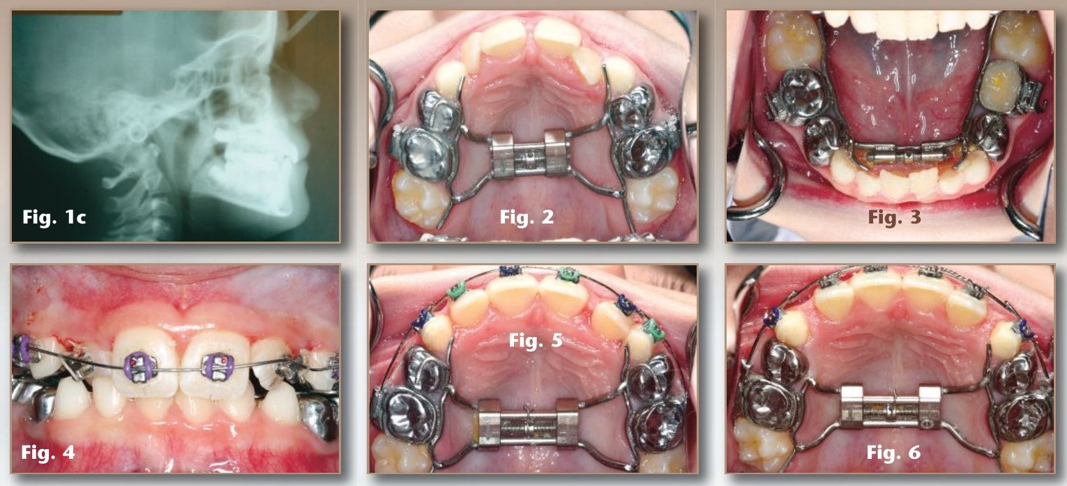
- Fig. 1c Pre-treatment cephalometric radiograph.
- Fig. 2 Upper orthopedic expander, banded to the E's; expansion after 1 month.
- Fig. 3 Lower orthopedic expander (Palex screw); expansion after 1 month.
- Fig. 4 Upper anterior brackets placed after transverse development.
- Fig. 5 Upper arch form slightly overcorrected.
- Fig. 6 Upper anterior space consolidated lateral to lateral.
In short, nothing good was going to happen for G.J., from this time forward, without intervention. Due to his retruded profile, thin lips, and obtuse nasio-labial angle, the treatment approach was geared toward non-extraction of permanent teeth. Goals were to increase arch width and to advance anterior teeth to increase arch length so all the permanent teeth had room to erupt in a near normal position.
Appliances were inserted on 2-18-09. Both upper and lower orthopedic expanders were cemented using GI cement. The appliances were banded to the E's (2nd primary molars) in both arches. Expansion screws were turned at a rate of one quarter turn every 4 days on both the upper and lower arch. Figs. 2 and 3 show expansion after 1 month. Expansion continued off and on and was monitored in both arches for approximately 6–8 months until lateral arch development was sufficient.
On average G.J. was seen every 4 weeks. Upper anterior brackets were placed after transverse development was proceeding in the upper arch (Fig. 4). Sagittal and transverse development continued in the upper arch until the arch form was slightly overcorrected (Fig. 5). The upper anterior space was consolidated from lateral to lateral (Fig. 6) with excess space mesial to the primary cuspids. Because the lower laterals were delayed in eruption, and because of lower anterior arch constriction, bracketing of the lower anteriors lagged behind the uppers. In the case of G.J., the lower primary cuspids and the permanent central incisors were initially bracketed while creating room for the laterals (Fig. 7), while the upper arch form was maintained. Continued bite opening and lateral development enabled bracketing of all lower permanent incisors (Fig. 8). The initial arch wires were 0.16 nickel titanium with small RM locks placed bilaterally against the mesial portion of the molar tubes on the anchor molars so the arch wire would extend in front of the anterior brackets by 1–2mm. By pushing the wire in the anterior bracket slots with the locks against the molar tubes an anterior force was delivered to the front teeth. The opposite occurred in the molar teeth with a distally directed force, but with anchorage in the posterior being greater, the net effect was advancement of the front teeth. The wire in the buccal segments bowed out reducing lateral lip pressures as well (Figs. 9 and 10). The upper arch wire sequence was 0.16 niti, 0.16 reverse curve niti, and 0.22 stainless steel. The lower arch wire sequence was 0.16 niti, and 0.16 stainless steel.
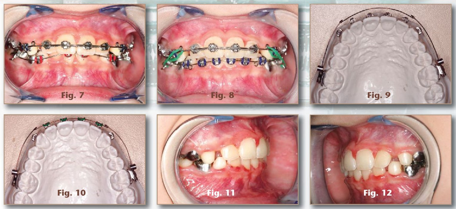
- Fig. 7 Lower primary cuspids and permanent central incisors bracketed while creating room for the laterals.
- Fig. 8 All lower permanent incisors bracketed.
- Figs. 9 and 10 RM locks against the molar tubes; archwire bowed out in the buccal segments.
- Figs. 11 and 12 Right and left buccal views immediately after appliance removal (Phase I, 18 months).
The total treatment time in the case of G.J. was 18 months for Phase I. Figures 11–12 show left and right buccal images taken immediately after appliances were removed. The post treatment panoramic and cephalometric x-rays are shown in Figs. 13 and 14. Note the space created for the second eruptive phase involving exchange of permanent cuspids and premolars in all quadrants. An upper removable Hawley and a banded lower lingual holding arch were used as retainers for G.J. These retainers were adjusted at intervals to allow eruption of the permanent premolars and cuspids. Figs. 15, 16, and 17 were taken 2, 10, and 22 months post treatment. Long term follow-up pictures 28½ months after phase I show the dental arches, the occlusion, and facial photographs (Figure 18). Note the arch symmetry during transition with ample space for all permanent teeth. Also note the profile, which is not too forward creating a bimaxillary protrusion, but is well balanced. This technique due to slow biologic arch development does not move the teeth off the ridge, or place them too far forward, but allows alveolar remodeling leaving the teeth well anchored in alveolar bone. After permanent teeth are erupted, a phase II with comprehensive bracketing will be accomplished if necessary.
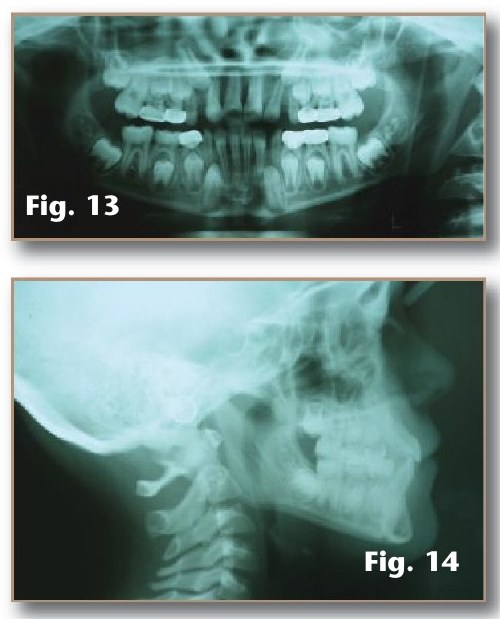
- Fig. 13 Post-treatment panoramic radiograph.
- Fig. 14 Post-treatment cephalometric radiograph.
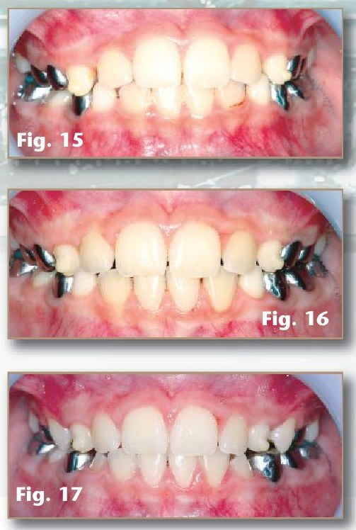
- Figs. 15, 16, 17 2, 10 and 22 months post-treatment.
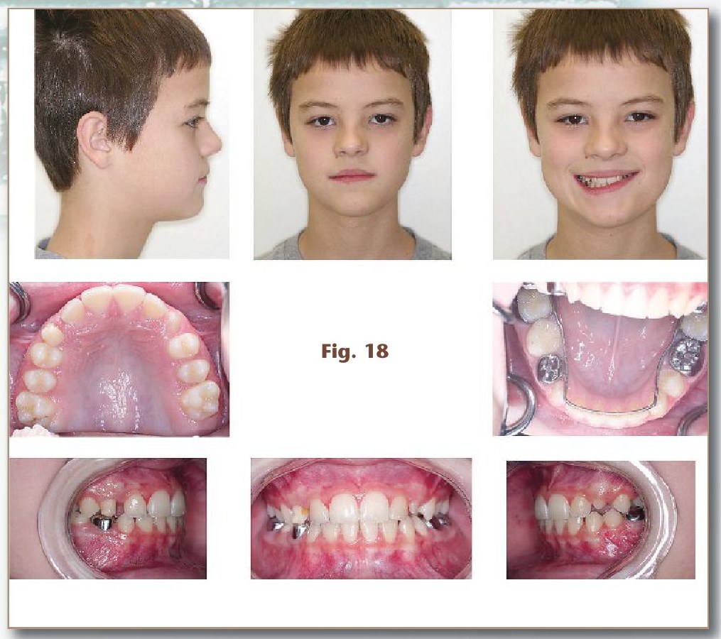
- Fig. 18 28½ months after Phase I: facial photographs, arches and occlusion.
Because the appliances in the dual arch orthopedic system are fixed (cemented) rather than removable, there is an added benefit of not having to manage patient cooperation for wearing the appliances. A major key in the dual arch orthopedic system is the anchor molars. Anchor molars are not the traditional first permanent molars (6's), but rather the second primary molars (E's) in both arches. Sufficient root development must be available on the second primary molar teeth, as these teeth must remain intact until the orthopedic expanders are removed. The design of the upper and lower orthopedic expanders with the second primary molars (E's) as anchor molars is shown in Figures 2, and 3. Note the lingual extensions on the appliances from the mesial of the primary cuspids (C's) to the distal of the permanent first molars (6's). Note also, the lower screw is smaller and is a palex screw. The posterior extensions allowed expansion of the permanent first molars in both arches simultaneously. The anterior extensions move the primary cuspids laterally as the appliance was turned. Expansion on either the upper or lower appliance can be discontinued for intervals to allow the opposing arch to catch up if posterior teeth are nearing a cross-bite. Thus, occlusal coordination can be maintained throughout treatment in the posterior quadrants, while the incisors are coupled anteriorly.
I learned to use the second primary molars as anchors or handles from Dr. Bill Hang, who learned from Dr. John Mew. Anchoring on the second primary molars allows treatment to commence when root development on newly erupted permanent first molars is not complete. Damage in the form of resorption or blunting to permanent first molar roots is minimized, and or avoided all together. In addition, because sufficient room is created in the dental arch, impactions are minimized. Treatment follows the natural growth direction of the jaws (down and forward), and thus, TMJ dysfunction is minimized or greatly reduced. Because arch development occurs both transversely and sagitally, (in an anterior direction), at the same time, the upper airway and nasal patency is improved. Thus, nasal respiration may now be possible in a child who was a mouth breather.
Arch width and arch length are adjusted for each individual child using the combination of the fixed expanders and brackets on the anterior teeth along with archwires. A second key for the dual orthopedic arch development system is the use of locks on the archwires mesial to the molar tubes. These locks act as stops and can be adjusted to advance the anterior teeth (See Figs. 9 and 10).
After twenty years of experience treating young children, dual arch orthopedics has proven to be an efficient and effective strategy for arch development prior to eruption of the full permanent dentition that provides both control and coordination of the dental arches. A simple analogy that I use to explain this concept to parents is that the mouth is a parking lot, and the teeth are the cars. Due to crowding, there is not enough room to park all the teeth in their correct spot. Thus, we treat to enlarge the parking lot (orthopedics) so that each tooth has a space to park, and this space is where that tooth needs to park to fit and function properly. By achieving this during the active eruptive phase, teeth will not require movement over large distances, enhancing long term stability. Treatment strategies are a continuum just as growth is a continuum, and thus any appliance system needs to be adaptable to the growing child. Applied correctly, and at the right time, dual arch orthopedics helps guide a developing malocclusion back to a state of normalcy.
The ideal time to start treatment is during, or just after transition of the upper and lower incisors. Maximal growth in the lateral width of the upper and lower jaws occurs with eruption of the upper centrals and lower laterals (29). It is also during this transitional period when the maximum arch length in the mouth is achieved. The anterior and posterior limits of the dentition are larger at this time than at any other time in the child's life. In fact, arch length decreases as the posterior primary teeth are replaced by permanent teeth. This is mainly due to the size difference between the primary second molars and the permanent second premolars which will eventually replace them (called E space, or leeway space). The important thing to remember about dental development and the exchange of teeth is that early crowding coincides with a period of rapid growth of the facial skeleton. Craniofacial growth is 80–90% complete by the age of twelve (18), when most traditional orthodontics is started on children. In addition, studies document no future increase in lower intercanine width occurs after incisor eruption for relief of crowding that might be present (30).
Crowding after the first transition (permanent incisors and permanent first molars) does not improve with continued growth or advancing age. Details of anterior incisor alignment during eruption and how to handle crowding is covered in detail in my previous article entitled "Early alignment of lower incisors; first thing first" (1). Knowing the normal growth and development of the child's dentition is critical in the decision of if and when to intercept and treat early.
In my practice, 85–90% of children will require phase II treatment, and the second phase on average is 9–12 months. The second phase is typically started with or soon after eruption of second permanent molars. Typically retainers (fixed lower lingual arch and an upper removable Hawley) are worn until the second phase (full braces) is started.
The mechanics described for the dual arch orthopedic system allow the teeth and jaws to move along natural growth vectors. The jaws are developed both transversely and forward, mimicking natural growth, which improves the posterior airway space behind the tongue, increases the nasal base, and allows more room for the tongue to assume a natural rest posture, all of which add to stability. Light continuous force induces bone remodeling in the alveolar portion of the jaws as the anterior teeth are moved orthodontically into their proper position and alignment. In the upper arch true expansion occurs, but in the lower alveolar remodeling occurs which allows the lower posterior teeth to upright. Coordinating both arches allows posterior occlusion to be maintained throughout treatment, and allows bite forces to be directed along the long axis of the molar teeth. In this way, narrow dental arches can be widened to accommodate the permanent teeth in the lower arch without the need for extraction.
Failure to intercept after the first transitional period, in which there is significant anterior crowding, will result in continued crowding, a worsening of the malocclusion, and it places the child at an increased risk for permanent tooth removal. Extraction of premolars to reduce crowding decreases arch length, thus effectively reducing the size of the maxilla and mandible (opposite of natural growth). This results in reduced space for the maxillary sinuses and nasal airway, which may lead to snoring, hypoxia, and obstructive sleep apnea later in life (13).
Early intervention is advantageous for improving facial balance, normalizing growth and dental development, improving esthetics, improving nasal patency and the patient's ability to breathe normally thru the nose, as well as lessening the need for removal of permanent teeth. In an article on the origins of malocclusions, the authors lend support for an early treatment approach that increases the volume of alveolar bone supporting the teeth and expanding the dental arches with orthodontics and dentofacial orthopedics during growth and development. Their research provides support for the development of orthodontic therapies that increase jaw dimensions rather than the use of tooth removal to relieve crowding (22). The philosophy of orthopedic arch development during the mixed dentition is also supported by Dr. John Mew, who creates beautiful balanced faces without the use of fixed appliances (21, 27).
Early in my career I had the privilege of attending a continuing education lecture on early treatment strategies from Dr. Ron Bell, an orthodontist. In one of his many articles he states: "The early mixed dentition (6 to 9 years of age) is a period highly prone to localized malocclusion factors that may result in severe problems if untreated. Interceptive orthodontics and guidance of eruption concepts elevate management of the developing occlusion to include recognition of factors producing a malocclusion and the implementation of treatment procedures to eliminate or minimize the effects of the malocclusion factors on the developing occlusion (8). The interventions reflect treatments designed to alter an existing abnormal situation, influence the eruption patterns and positioning of the permanent teeth during transition, and create an optimum potential for normal occlusion development for the individual child. There are two major factors that generally influence the delicate balance of facial growth; the airway and the manner in which the tongue functions (31). Early correction allows treatments that are less complex, less time consuming, and more physiologically tolerable than demands in adolescent and adult patients (30). The long term benefits of early treatment directed toward more harmonious occlusion may eliminate or minimize deleterious anatomical and functional growth factors (32).
In my office, case selection for dual arch orthopedics is heavily dependant on soft tissue features such as profile, lip thickness, and nasio-labial angle, as well as the total discrepancy in the mixed dentition and orthodontic analyses. Treatment is typically limited to cases with total discrepancies 5.0mm or less. With E-space intact, significant crowding in the 7–8mm range can be resolved using this technique. In addition, posterior airway space, size of tonsils and adenoids, snoring, mouth breathing, allergies, habits, and rest oral posture must all be factored in the overall treatment plan. Obviously, patient cooperation is essential, and of course parents have to be on board as well.
In this article I have described the dual arch orthopedic technique that I currently use for early orthopedic/orthodontic cases in my office. I have shown a detailed case (G.J.) from start to finish for this phase as a typical example of the treatment results, as well as given detailed description of the appliance design and purpose. In addition, I have given indications, supported in the literature, for early arch development in the growing child. Look for these in your own patients. Providing this type of treatment is very rewarding for both the patient and the dentist!
References
The numbered citations (1–32) appear in the text as printed. The Journal did not print the reference list with this article.